
Mohs laboratory plays a critical role in the field of dermatology and skin cancer treatment. As skin cancer rates continue rising, Mohs surgery has become an invaluable treatment approach.
The specialized Mohs laboratory allows for precise microscopic analysis of cancerous skin tissues, enabling targeted removal of tumors while preserving healthy tissue. This results in improved cure rates and cosmetic outcomes. In this article, we will explore what happens inside a Mohs lab and why it is integral to skin cancer surgery.
Mohs surgery, named after Dr. Frederic Mohs, is a highly effective technique for treating common skin cancers like basal cell carcinoma and squamous cell carcinoma. It is also sometimes used to treat rare cancers like melanoma.
The key advantage of Mohs surgery is its method of surgically removing cancerous tissues layer by layer while assessing each layer microscopically. This allows surgeons to precisely map and eradicate tumors while minimizing damage to healthy tissue. Traditional surgical excision is less exact, often removing large areas of healthy skin to ensure full tumor removal.
During Mohs surgery, a dermatologic surgeon first surgically exises visible cancerous tissues. These samples are mapped, labeled, and transported to the Mohs laboratory. Here, a specialized technician processes and microscopically analyzes the tissue samples, looking for any remaining cancer cells. If found, their exact locations are mapped, and the surgeon uses this information to surgically remove additional tissue layers precisely where cancer cells persist.
This cycle continues until no more cancer cells are detected microscopically. This unique ability to pinpoint and remove 100% of cancer sets Mohs surgery apart. It results in up to a 99% cure rate for common skin cancers while maximizing preservation of healthy tissue.
Specialized Mohs laboratories contain unique equipment tailored to facilitate tissue analysis during Mohs surgery. While Mohs labs vary, most contain similar key components.
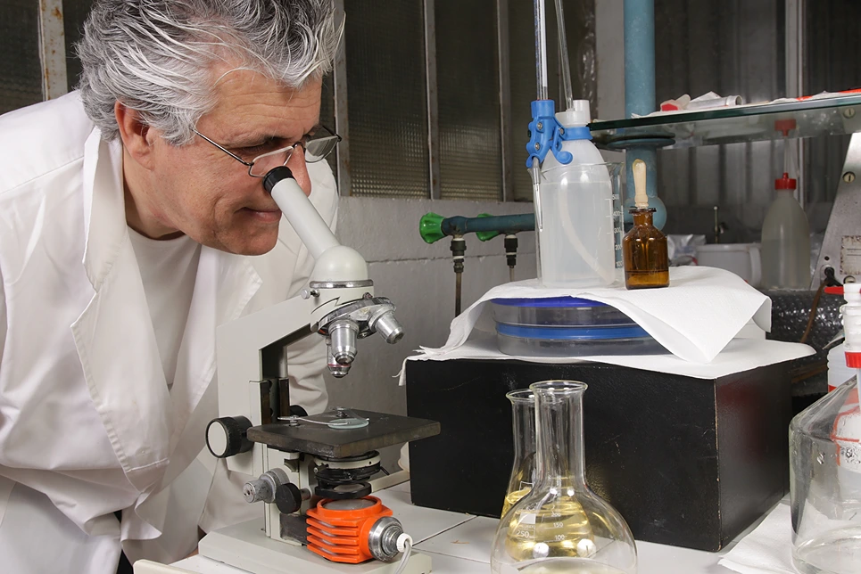
At the core is the surgical microscope connected to a large computer monitor. This enables the lab technician to minutely examine tissue architecture and cytology to detect any lingering malignant cells. Sophisticated lighting and camera equipment connected to the microscope produce high-resolution images used for precision mapping.
The lab contains cryostats which freeze tissue samples for cutting thin slices for microscopic mounting and immunohistochemical staining. Staining makes cancerous cells more visible. Common stains used include hematoxylin and eosin.
Other essential lab equipment includes microtomes for precision tissue slicing, grossing stations for initial tissue preparation, special fixatives and stains, and storage for frozen tissue blocks. Pathology laboratories also contain fume hoods, biohazard safety equipment, and sanitization tools to maintain a sterile environment.
Digital pathology and imaging systems allow technicians to digitally scan and annotate slides for surgical mapping. Accurate labeling and documentation are critical for tracking the excised tissues' precise locations on the patient.
Overall, assembling an effective Mohs laboratory requires specialized medical expertise and significant financial investments of $100,000 or more. Ongoing costs include lab supplies, reagents, tissue processing materials, and highly trained staff.
On the day of surgery, the patient area is first sterilized, and local anesthetic administered for patient comfort. The visible tumor is excised with careful margins using a sterile scalpel or curette. Hemostasis is achieved using cautery.
The surgeon then precisely maps, labels, and orients the tissue sample before transferring it to the Mohs laboratory. Strict handling protocols preserve tissue architecture and orientation.
Samples are sectioned, processed, and frozen rapidly to optimally visualize cancer cells.
In the lab, the frozen tissue block is sliced to produce thin sections which are mounted on microscope slides and stained. The technician systematically scans the slide under the microscope, analyzing architectural and cytologic features to detect any residual cancer cells.
If cancer cells are found, the technician maps their exact locations using annotations and digital images. This map is delivered to the surgeon, guiding precise excision of additional tissue layers only where cancer persists.
This cycle repeats until no cancer cells remain. Patients may undergo one to three stages depending on the extent of the tumor. After clear margins are achieved, the surgical wound is closed or reconstructed as needed.
Patient safety and comfort are paramount throughout Mohs surgery. Vital signs, bleeding, and pain levels are closely monitored. Local anesthetics manage pain while preserving consciousness. Antibiotics and other medicines prevent complications like infections.

Pathological examination is the cornerstone of Mohs surgery. Accurate frozen section analysis and mapping of cancer cells enables the targeted layer-by-layer removal which underlies Mohs' effectiveness.
During surgery, the Mohs technician employs specialized techniques to process and stain tissue samples. Freezing, sectioning, and staining must be carefully conducted to avoid distorting delicate dermatopathologic features.
The technician thoroughly analyzes stained sections under high-power microscopes. They must recognize cancer cell morphology and patterns to pinpoint residual tumors. Knowledge of skin histology is essential for distinguishing benign tissues from malignancy.
The technician carefully maps and documents the slide to guide the surgeon in removing additional layers. Precise labeling, photography, and face-to-face communication ensure clear transfer of information.
Mohs laboratories require diligent tissue processing protocols. Frozen tissue blocks must be stored carefully indexed by patient and anatomical origin. Frequent quality control ensures optimal stain quality and microscope function.
By enabling complete and targeted removal of tumors, Mohs surgery offers patients the highest cure rates of any skin cancer treatment. Studies show Mohs achieves a 99% 5-year cure rate for common skin cancers.
This compares favorably to cure rates of 85-95% for standard excision. By removing less healthy tissue, Mohs also improves cosmetic outcomes and reduces complications like infections and pain.
Patients undergoing Mohs surgery can expect minimal scarring since mostly diseased tissue is excised. Wound size depends on the cancer's size and location. Small wounds may heal by secondary intention while larger ones require surgical reconstruction using skin flaps or grafts.
Healing typically takes several weeks during which patients must keep the site clean and bandaged. Stitches are removed after 1-2 weeks. Doctors closely follow-up to monitor recovery and watch for recurrence.
Follow-up visits include physical examination of the surgical site and scar. Photographic documentation is helpful for comparison. Patients may need occupational or physical therapy to improve function or range of motion if muscles or nerves were affected.
Overall, Mohs surgery provides patients the most effective and precise skin cancer treatment available. When combined with skilled reconstruction, it optimizes cure rates, aesthetics, and quality of life - a testament to the vital role of the Mohs laboratory.

Mohs laboratory is an indispensable component of modern skin cancer care. By facilitating microscopic analysis of excised tissues and pinpoint mapping of residual cancer, the Mohs lab enables unparalleled surgical precision and effectiveness.
Skilled dermatologists, surgeons, and technologists work in tandem with specialized equipment to eradicate tumors layer-by-layer while maximizing healthy tissue preservation.
This interdisciplinary approach sets Mohs surgery apart, leading to improved patient prognoses, aesthetics, and quality of life. As skin cancer rates increase, the vital role of the Mohs laboratory will only continue growing.
If your desired appointment type or preferred provider is unavailable online, kindly call (978) 525-0100 for Peabody, MA and (603) 742-5556 for all New Hampshire locations. Alternatively please feel free to send us your request via the patient portal, or via email at info@dermskinhealth.com
*For medical dermatology appointments in MA please dial (978) 525-0100 or fill out the appointment request form above.