
Spotting a new or changing lesion on your skin can be concerning. It’s only right to get it evaluated quickly by an expert dermatologist to determine if it's harmless or requires treatment. Here at Dermatology and Skin Health, we offer a range of advanced techniques to remove benign growths and treat precancerous or cancerous skin lesions.
Read on to explain the key differences between common options like excision, curettage, electrodesiccation, cryosurgery, and Mohs surgery. This is so you’ll understand the optimal treatment approach based on factors like the type of lesion, size, location, risks, and your goals for removal.
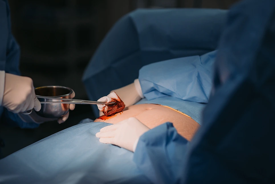
Excision involves surgically cutting out the entire lesion with a scalpel or scissors, including a margin of normal-looking skin around it. This is commonly done for growths that appear abnormal or have an uncertain diagnosis, allowing the tissue to be sent for biopsy after removal. Excision is the most invasive approach but offers the best chance of complete removal.
Curettage uses a special surgical instrument called a curette, which has a scoop or ring shape on the end. This is used to essentially scrape away the lesion down to the deeper skin layer called the dermis. Curettage is less invasive than excision and often used for actinic keratoses and superficial basal cell carcinomas.
Electrodesiccation utilizes an electrical current to heat and desiccate or dry out lesion cells, causing them to die and slough off. It’s applied to the lesion site after curettage to control bleeding and destroy any remaining abnormal tissue. This is suitable for very thin, superficial lesions.
In Mohs surgery, thin horizontal layers of skin are incrementally removed and immediately examined under a microscope by the surgeon. Additional layers are excised and reviewed until no evidence of tumor cells remains. This staged approach allows maximal preservation of healthy tissue.
Cryosurgery involves freezing lesions using liquid nitrogen. The extreme cold temperature damages cells, causing the lesion to slough off. It’s less invasive than other techniques and often used for small, low-risk growths.
| Treatment | Invasiveness | Purpose | Suitability for Different Types of Lesions | Complications |
| Excision | Most invasive | To remove the entire lesion, including a margin of normal-looking skin around it | Commonly used for growths that appear abnormal or have an uncertain diagnosis | Scarring, pain, bleeding, wound breakdown, infection, insufficient margins, and nerve damage |
| Curettage | Less invasive than excision | To scrape away the lesion down to the deeper skin layer (dermis) | Often used for actinic keratoses and superficial basal cell carcinomas | May generate more unaesthetic scars compared to conventional surgical procedures |
| Electrodesiccation | Less invasive than excision | To heat and desiccate or dry out lesion cells, causing them to die and slough off | Suitable for very thin, superficial lesions | Scarring is common |
| Mohs Surgery | Invasive, but less tissue damage compared to excision | To remove thin horizontal layers of skin incrementally until no evidence of tumor cells remain | Ideal for lesions caused by skin cancer | Scarring is also common |
| Cryosurgery | Least invasive | To freeze lesions using liquid nitrogen, causing the lesion to slough off | Often used for small, low-risk growths | Dyspigmentation, hair loss, and tissue distortion |
Careful evaluation of any suspicious skin lesion is critical prior to determining the best treatment approach. At Dermatology and Skin Health, our expert dermatologists utilize visual examination techniques, dermoscopy, and skin biopsy when warranted to fully assess lesions before intervening.
Visual examination involves carefully looking at the lesion for the “ABCDEs” of melanoma—asymmetry, irregular borders, varied colors, large diameter, and evolution or changes over time. Other concerning features include bleeding, crusting, and changes in sensation.
Dermoscopy lets us examine lesions under high magnification. Certain patterns and vascular structures provide clues to differentiate between benign, precancerous, and cancerous lesions.
While many lesions have a clear clinical diagnosis, a biopsy is sometimes needed to confirm. This involves numbing the area and using a punch tool to extract a small sample of the lesion for microscopic analysis.
In determining treatment, we consider the lesion’s size, location, thickness, and potential risks. Treatment goals are also important—is complete removal necessary or is preserving surrounding tissues a priority?
A cross-sectional study conducted at Brigham and Women's Hospital Dermatology in 2018 found that patients prioritize different aspects of the treatment experience based on factors such as age, gender, race, insurance status, and history of malignancy. Health and preferences also factor into the decision.

At Dermatology and Skin Health, surgical excision is our preferred treatment for certain types of concerning skin lesions, including:
Lesions that appear clinically suspicious for melanoma. Excision allows for biopsy and staging if melanoma is confirmed. A systematic review published by NCBI in 2019 states that surgical excision carries the highest cure rates for all skin cancers and is the first-line treatment for melanomas and high-risk nonmelanoma cancers.
Large moles or nevi in areas of friction and irritation, such as the back, neck, and abdomen. Removing unstable nevi prevents progression to melanoma. A 2013 study on the optimal management of common acquired melanocytic nevi suggests that surgical excision is a common treatment for these skin lesions. The study also mentions that the aim of cosmetic surgery is largely the removal of the visible lesion.
Ulcerated lesions that won’t heal, as these can hide a cancerous process. A 2009 study on excision of pre-ulcerative forms of Buruli ulcer disease found that bacilli may extend beyond the actual size of the lesion, supporting the need for surgical excision.
Excision is also preferred when the conservation of the surrounding healthy skin and tissues is not a priority, such as on the back. The procedure allows for definitive removal and comprehensive pathological analysis.
Curettage involves using a sharp, spoon-shaped instrument called a curette to selectively scrape away skin lesions in a meticulous manner. At Dermatology and Skin Health, we frequently use curettage to remove actinic keratoses and certain shallow skin cancers like superficial basal cell carcinomas.
According to the Mayo Clinic, the process begins by cleansing and numbing the treatment area with a local anesthetic. The curette is then used to scrape off the damaged cells. This may be followed by electrosurgery, where a pencil-shaped instrument is used to cut and destroy the affected tissue with an electric current.
For raised or thicker lesions, we may do several repeated scrape passes to fully remove tissue down to normal skin levels. Bleeding is controlled with electrodessication. The procedure leaves behind a shallow, clean wound bed that can then heal by secondary intention or be treated with adjunctive therapies.
Curettage is advantageous for removing thin, scaly precancerous lesions on the face, hands, and other cosmetically sensitive areas. It preserves the deeper skin layers, allowing excellent healing and cosmetic results.
After curettage, electrodessication can help control bleeding while destroying any remaining abnormal cells in the lesion site. At Dermatology and Skin Health, we use hyfrecation, which applies high-frequency electrical current through a fine needle tip placed on the wound bed.
The treated area develops a dry eschar or scab within 24 hours. The scab naturally sloughs off over the next 2-4 weeks as new skin cells replace it. Keeping the area moist with ointment prevents crusting and speeds healing.
Mild pain, redness, and swelling are common for a few days following electrodesiccation treatment. Patients can take over-the-counter pain medication as needed for discomfort. Some temporary numbness or nerve sensitivity in the area can also occur.
Proper wound care is vital - you’ll need to gently cleanse the site daily and apply antimicrobial ointment until healed. Avoid picking at scabs. Watch for signs of infection like increased pain, swelling, oozing, and redness.
Once healed, the skin may remain slightly lighter in color, but little scarring occurs since the deeper layers remain intact. Avoid direct sun exposure. Let us know if the treated area does not fully heal within 4 weeks, as this may indicate recurrence.
While electrodesiccation is highly effective for eradicating many superficial skin lesions, it does have some limitations depending on the characteristics of the growth.
A 2019 study found that electrodesiccation was effective in treating seborrheic keratoses, with average lesion sizes around 8.8mm - within the 1cm thickness suitable for the technique according to the passage. However, deeper nodular growths with roots extending far below the skin surface are harder to fully desiccate down to the base, increasing chances of recurrence.
A 2022 study conducted at the Department of Infectology, Dermatology, Diagnostic Imaging and Radiotherapy in Brazil found that curettage and electrocoagulation, which includes electrodesiccation, had comparable local recurrence rates to conventional surgery for low-risk basal cell carcinoma lesions.
Very large or extensive lesions are also difficult to treat, as prolonged heating can damage healthy surrounding tissues. Lesions larger than 2cm in diameter often require staged treatments.
The technique is ideal for eliminating actinic keratoses, the precancerous scaly patches caused by sun exposure. It also excels at removing benign viral warts and shallow, low-risk basal cell carcinomas.
For suspected melanomas or atypical moles, excisional biopsy is recommended over electrodesiccation since it allows pathological analysis of lesion depth and margins.
A 2022 article in StatPearls recommends performing an excisional biopsy on suggestive melanoma lesions so a pathologist can confirm the diagnosis, aligning with the passage's advice.
In summary, electrodesiccation is a valuable technique for treating many common superficial skin lesions. But other approaches like surgery may be preferable for deep, large, or high-risk lesions requiring microscopic examination.
Mohs micrographic surgery offers the highest cure rate for high-risk skin cancers located on the head, neck, hands, feet, and other cosmetically sensitive areas where tissue preservation is vital. At Dermatology and Skin Health, Dr. Mendese performs Mohs surgery for complex or recurrent cases. Here's an overview:
First, after numbing the area, the visible tumor is debulked and a thin layer of tissue is removed. This layer is color-coded and oriented for microscopic mapping, then frozen and processed while the patient waits.
The surgeon meticulously examines the tissue under a microscope, looking for any remaining cancer cells at the periphery and base. If positive margins remain, the next incremental layer is precisely removed from the involved area and examined.
This process continues layer-by-layer until the margins are completely clear of cancerous cells. This allows maximal preservation of healthy tissue surrounding the tumor. Once clear margins are achieved, the resulting surgical defect can be allowed to heal by second intention or reconstructed.
Reconstruction options include local tissue flaps, skin grafts from elsewhere on the body, or closure by layering together skin edges. Mohs surgery typically takes several hours to complete but is performed in the outpatient setting with local anesthesia.
While Mohs micrographic surgery offers unmatched precision in eradicating skin cancers, there are some risks and potential complications to consider:
The lengthy, meticulous process requires specialized surgical training and skill. So choosing an experienced, high-volume Mohs surgeon is vital to minimize risks.
Mohs surgery is typically an outpatient procedure performed under local anesthesia. But extensive reconstruction may require a short hospital stay for recovery.
While Mohs surgery carries some risks like any invasive procedure, our expert surgeons minimize complications through precision technique while maximizing cancer removal. Letting us know your complete medical history allows us to safely perform Mohs surgery when indicated.
Here at Dermatology and Skin Health, we frequently use cryosurgery to treat certain benign and malignant skin lesions, especially when preservation of surrounding healthy tissue is a priority. Some common indications include:
A 2015 retrospective study conducted in Turkey found that 95.92% of patients had a clinical diagnosis prior to cryosurgery treatment.
Cryosurgery is not recommended for suspected melanomas or atypical moles, which require excisional biopsy for accurate diagnosis and staging. Let us know if you notice any spot on your skin with concerning features so we can determine if cryosurgery is appropriate.
Cryosurgery offers multiple benefits that make it an appealing option for treating certain benign and malignant skin lesions:
In summary, cryosurgery should be considered for small, superficial skin lesions when preservation of function and aesthetics are a priority. The minimal invasion and simple recovery make it an ideal option for many patients.

If you notice any new or changing spots on your skin, schedule an appointment with the experts at Dermatology and Skin Health. We will examine your skin carefully, order diagnostic tests if needed, and diagnose benign growths versus suspicious or cancerous lesions.
Based on the type of lesion, location, depth, and your goals for treatment, we can advise on the best option - whether that's actively monitoring small lesions, using non-invasive approaches like topicals or cryosurgery for low-risk growths, or surgical excision for aggressive or malignant tumors.
Our specialized Mohs surgery service excels at eradicating high-risk cancers on the face, head, neck, hands, and other functionally important areas where preserving healthy tissue is vital. You can trust our fellowship-trained Mohs surgeon, Dr. Mendese, to achieve clear margins and optimal cosmetic results.
If your desired appointment type or preferred provider is unavailable online, kindly call (978) 525-0100 for Peabody, MA and (603) 742-5556 for all New Hampshire locations. Alternatively please feel free to send us your request via the patient portal, or via email at info@dermskinhealth.com
*For medical dermatology appointments in MA please dial (978) 525-0100 or fill out the appointment request form above.