
Losing part of your ear to skin cancer can be devastating.
Beyond the emotional impact, physical changes to this complex cartilaginous structure can affect not only appearance but also hearing and function. Fortunately, with the right surgical approach, skilled reconstructive techniques can restore the natural contours and symmetry of the ear after skin cancer removal.
Ear reconstruction following Mohs surgery represents a complex process, but also an opportunity. In the hands of experienced reconstructive surgeons, this restorative work aims to rebuild the structural framework of the ear and recreate natural aesthetic form. Utilizing a combination of skin flaps, cartilage grafts, and meticulous layering approaches, post-Mohs ear repair can produce excellent functional and cosmetic outcomes.
Mohs micrographic surgery has become a common technique for removing different skin cancer types on the ears, including basal cell and squamous cell carcinomas. By progressively excising and examining cancerous tissue under a microscope, Mohs surgery aims to remove tumors while conserving as much healthy tissue as possible.
However, the intricate cartilaginous anatomy of the ear means skin cancer removal can create complex defects. The ear’s unique topography—with many ridges, depressions, and a thin covering of skin over cartilage—poses challenges for reconstruction after Mohs surgery.
Depending on cancer size and location, the Mohs procedure may require excising skin, cartilage, or both. This can leave noticeable dents, holes, or malformed areas. In addition to cosmetic concerns over the ear’s appearance, this type of tissue removal can also affect structure and function.
That’s where reconstructive surgery comes in. The goals of ear reconstruction after Mohs surgery include:
With careful planning and execution, ear reconstruction can successfully meet these goals following skin cancer removal.
Ear defects after Mohs surgery can vary considerably depending on:
To address this spectrum, plastic and reconstructive surgeons have a range of repair techniques from which to choose:
| Technique | Description | Advantages | Disadvantages |
| Skin grafts | Use skin from behind ear or scalp | Less invasive, quicker | Less blood supply, need for second site |
| Local skin flaps | Rotate nearby skin into defect | Robust blood supply | More complex, longer surgery |
| Cartilage grafts | Graft ear/rib cartilage | Provides structure | Donor site, shaping challenges |
| Primary closure | Suture wound edges together | Simple, single site | Only for smaller defects |
The specific approach depends on the characteristics of the Mohs defect and patient factors like age and medical status. In many cases, a combination of modalities is used.
For example, a large defect on the ear's bowl involving loss of cartilage may require a skin flap for coverage along with a structural cartilage graft. Accurately assessing the post-Mohs defect and thoughtfully selecting from available reconstructive techniques is key to success.
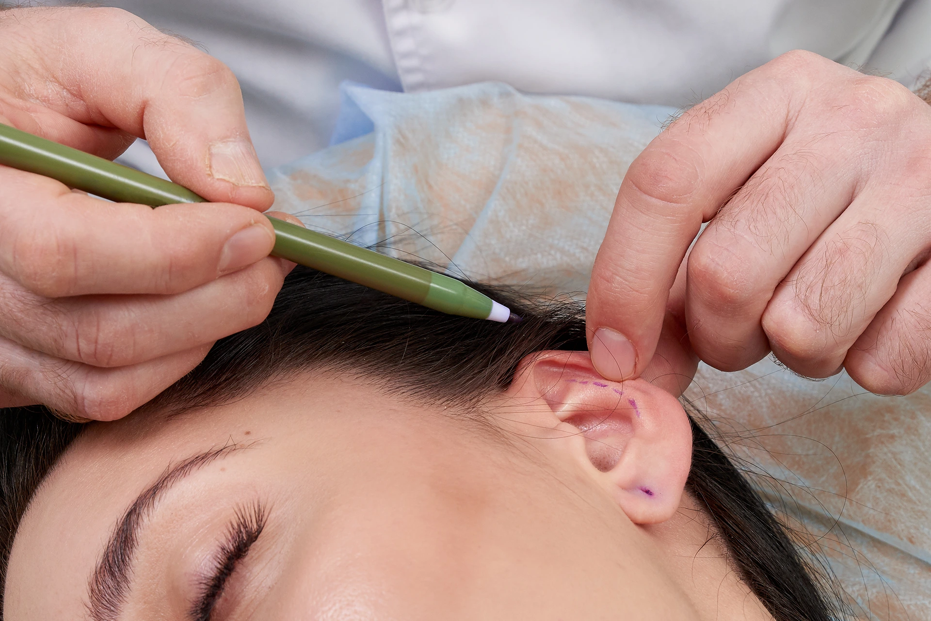
Meticulous surgical planning and execution forms the foundation of ear reconstruction after Mohs surgery. The main procedural steps include:
Throughout reconstructive ear surgery, maintaining adequate blood supply and venous outflow are crucial elements. Avoiding tension or distortion of the intricate ear structures also helps promote optimal healing.
For larger or complex defects, staging procedures over multiple surgeries may be required.
The recovery process after ear reconstruction aims to protect repairs while allowing optimal healing. Here’s an overview of what to expect:
Reconstructive surgeons will closely follow progress during the recovery period through frequent office visits and oversight. Patients play an important role by carefully following post-op protocols and reporting any worrisome symptoms.

Meticulous surgical planning and execution forms the foundation of ear reconstruction after Mohs surgery. The main procedural steps include:
Throughout reconstructive ear surgery, maintaining adequate blood supply and venous outflow are crucial elements. Avoiding tension or distortion of the intricate ear structures also helps promote optimal healing.
For larger or complex defects, staging procedures over multiple surgeries may be required.

The recovery process after ear reconstruction aims to protect repairs while allowing optimal healing. Here’s an overview of what to expect:
Reconstructive surgeons will closely follow progress during the recovery period through frequent office visits and oversight. Patients play an important role by carefully following post-op protocols and reporting any worrisome symptoms.
Precision Treatment for Select Skin-Based Breast Tumors Have you heard of Mohs surgery? You may…
Read MoreYour Complete Post-Mohs Recovery Plan After mohs surgery for skin cancer, you will need follow-up…
Read MoreMohs surgery offers precise removal of early-stage melanomas, preserving healthy tissue, but its use is…
Read MoreLearn about the healing process, recovery time, and essential care instructions following Mohs surgery on…
Read MoreEmpowering You with Knowledge on the Most Advanced Skin Cancer Treatment Mohs surgery removes skin…
Read MoreRefining the Skin's Surface for a More Even Appearance Dermabrasion after Mohs surgery can improve…
Read MoreThe One-Two Punch Against Skin Cancer: Mohs Surgery + Laser Therapy Laser surgery and Mohs…
Read MoreDiscover the surprising reasons why your body is drained and how to regain your energy.…
Read MoreShould You Take Antibiotics After This Common Skin Cancer Treatment? We Break Down the Pros…
Read More
If your desired appointment type or preferred provider is unavailable online, kindly call (978) 525-0100 for Peabody, MA and (603) 742-5556 for all New Hampshire locations. Alternatively please feel free to send us your request via the patient portal, or via email at info@dermskinhealth.com
*For medical dermatology appointments in MA please dial (978) 525-0100 or fill out the appointment request form above.