

Mohs surgery tools allow trained doctors to precisely remove skin cancer layer-by-layer. The specialized scalpels, curettes, forceps, and microscopes used in Mohs surgery help maximize removal of cancer cells while saving healthy skin.
In the fight against skin cancer, Mohs surgery serves as a tactical strike force uniquely equipped to seek and destroy tumors with pinpoint accuracy.
Using specialized surgical tools and iterative excision paired with microscopic analysis, Mohs surgeons can precisely eliminate cancerous skin layer by layer. This controlled, methodical technique made possible by optimized Mohs instruments achieves cure rates over 95% while maximizing preservation of healthy tissue.
In this guide, we’ll cover the essential tools that enable the unmatched precision of Mohs surgery. Gain insight into how skilled surgeons leveraging microscopes, scalpels, curettes and other Mohs tools can definitively conquer skin cancer while avoiding disfigurement.
Mohs surgery, named after Dr. Frederic Mohs, who developed it in the 1930s, is a specialized technique for removing certain common types of skin cancer, primarily basal cell carcinoma (BCC) and squamous cell carcinoma (SCC).
This procedure is distinct from standard surgical excision because the removed cancerous tissue is immediately examined under a microscope. This real-time analysis allows the surgeon to confirm whether all cancerous cells have been successfully removed while preserving as much healthy tissue as possible.
Several factors contribute to the precision of Mohs surgery:
The ability to microscopically examine excised tissue is indeed the key advantage of Mohs surgery, ensuring that no cancer cells are left behind.

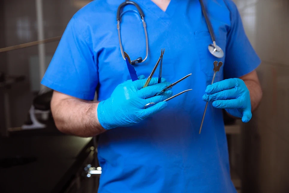
Mohs surgery requires a unique set of surgical instruments to remove tissue layers precisely and examine samples microscopically. Here are the key tools used:
Two main types of microscopes are used in Mohs surgery:
After each stage of excision, the tissue samples are meticulously prepared, stained, and examined under both microscope types to identify any remaining cancer cells.
Scalpels and blades are used to precisely excise the tissue layers. Common types include:
Our surgeon selects the appropriate blade for each excision stage, allowing controlled and incremental tissue removal.
Curettes are instruments with oval-shaped tips used for tissue scraping and removal. Mohs surgeons use curettes in all stages of tumor removal. Types used include:
Curettes provide precise control when removing tissue layers and when scoping the margins after tumor removal.
Forceps grasp and maneuver the tissue during excision. Common forceps used in Mohs surgery include:
Forceps enable our surgeon to orient, transfer, and flatten out tissue specimens for microscopic analysis.
Additional specialized tools used in Mohs surgery include:

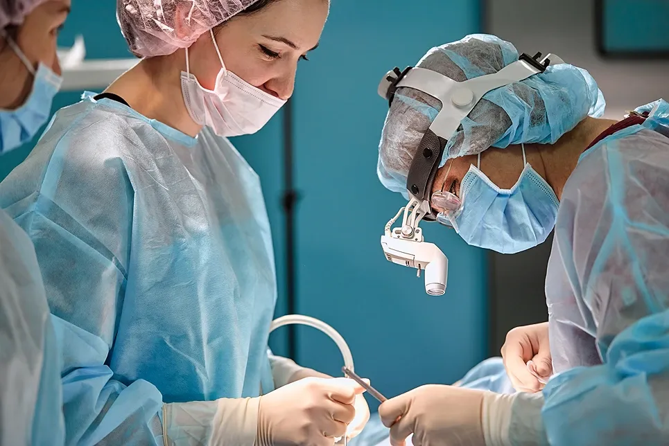
Our surgeons begin by administering local anesthesia to numb the area. Then, they use a scalpel, curette, or razor to remove the visible tumor along with a thin layer of surrounding tissue. Forceps assist in handling the excised tissue. This first stage aims to remove the obvious cancer cells.
The excised tissue samples are carefully cut, marked, and mapped. They are then processed for microscopic examination. This involves freezing the tissue and preparing it into thin sections for analysis.
The prepared slides of tissue are analyzed under both a stereomicroscope and a compound microscope to search for any remaining cancer cells. This step is crucial for determining if further excision is necessary.
If cancer cells are detected in the microscopic examination, our surgeons mark their exact location on a surgical map created during the initial excision. This mapping is essential for accurately identifying where additional tissue needs to be removed.
Using the precise mapping from the previous step, our surgeons return to the marked areas to remove additional tissue only from locations still containing cancer. This cycle of excision and examination continues until no cancer cells remain in the surgical margins.
Once all cancerous cells have been cleared, they close the wound using sutures or other methods. The entire process may take several hours, often under four hours for a typical Mohs surgery session.

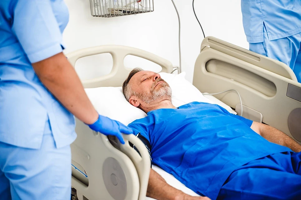
Recovery time after Mohs surgery varies based on factors such as the size and location of the removed cancer. Here’s what to expect during the recovery process.
Mohs surgery relies on an array of specialized surgical instruments to facilitate the controlled, incremental removal of skin cancer tissue. From microscopic analysis to meticulous excision, the tools utilized in Mohs surgery enable its precision and effectiveness.
Scalpels, curettes, forceps, and microscopes all play critical roles in the multi-stage excision and examination process. The surgeon utilizes these instruments to systematically identify and eliminate every cancer cell while protecting healthy tissue. It is this tool-dependent, layer-by-layer technique that makes Mohs surgery so highly effective.
Without the steady hands of trained surgeons and the purpose-built tools that enable the Mohs technique, this type of precision cancer removal would not be possible. While Mohs surgery requires advanced expertise and special equipment, its tissue-sparing precision leads to exceptional cure rates, excellent cosmetic outcomes, and enhanced quality of life for skin cancer patients.
If you have any suspicious growths or skin changes, see an expert dermatologist from Dermatology & Skin Health promptly for an exam. They can determine if Mohs surgery and its associated instrumentation is the optimal treatment approach.
[Don't delay - early detection and specialized Mohs tools together offer the best opportunity to conquer skin cancer. Contact us today.]
If your desired appointment type or preferred provider is unavailable online, kindly call (978) 525-0100 for Peabody, MA and (603) 742-5556 for all New Hampshire locations. Alternatively please feel free to send us your request via the patient portal, or via email at info@dermskinhealth.com
*For medical dermatology appointments in MA please dial (978) 525-0100 or fill out the appointment request form above.