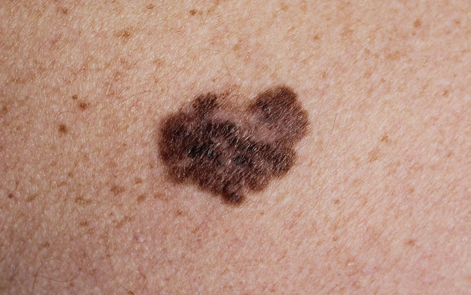

Mohs surgery has become a highly effective technique for removing certain common skin cancers while maximizing preservation of healthy tissue. However, Mohs is generally not considered the preferred treatment approach for melanoma, the most serious form of skin cancer.
Mohs micrographic surgery involves surgically removing skin cancer layer by layer, with each freshly excised tissue layer carefully mapped and examined under a microscope by the surgeon. This process allows the detection of even microscopic cancer cell extensions, enabling complete removal of the tumor.
Due to its ability to trace out borders with precision, Mohs surgery is often used to treat basal cell carcinoma and squamous cell carcinoma, two very common types of skin cancer. However, the role of Mohs surgery in treating melanoma is more limited.
Melanoma originates from pigment-producing cells called melanocytes and can quickly metastasize through blood vessels and lymphatics. Melanoma is staged based on its depth of penetration into layers of the skin, with higher stage tumors carrying greater risks.
Even in early stages, melanoma can be more aggressive and prone to spreading than basal or squamous cell carcinomas. Determining the true depth and borders of a melanoma lesion prior to removal can also be challenging.
Due to the tendency of melanoma to rapidly spread, a wide and deep surgical excision is recommended to remove the entire tumor with clear margins. In addition, a sentinel lymph node biopsy is often needed to determine if the melanoma has reached nearby lymph nodes.
Conventional excisional surgery allows removal of deeper portions of thicker melanoma tumors. Mohs surgery is typically limited to treating superficial skin layers and may not adequately excise deeper melanoma extensions.
Mohs’ layer-by-layer approach also makes complete lymph node staging difficult. For these reasons, standard surgical excision and lymph node dissection remains the gold standard for melanoma removal and staging.

In very early stage or lentigo maligna melanoma confined to the surface of the skin, Mohs surgery may rarely play a role. However, many dermatologists avoid Mohs even for superficial melanoma due to risks the procedure may miss portions of spreading melanoma.
In special cases like melanoma of the face, Mohs may be considered due to better cosmetic outcomes. However, conventional surgery is still typically preferred, and any Mohs surgery for melanoma requires very close follow-up monitoring.
With its propensity to spread, melanoma can often extend deeper than is detected on initial examination. If used in these cases, Mohs surgery runs a substantial risk of failing to remove the entire tumor, increasing chances the melanoma will recur.
Mohs’ limited depth of excision can also prevent accurate staging of the melanoma and assessment of lymph nodes. Attempting to use Mohs surgery to treat melanoma that has already advanced risks leaving cancerous cells behind.
Depending on the stage and progression of the melanoma, surgery may be accompanied by lymph node dissection, chemotherapy, immunotherapy drugs, or radiation therapy. This multidisciplinary approach maximizes chances of fully eradicating the cancer.
Research has also led to exciting new targeted drug therapies that are improving prognosis and survival rates even for advanced melanomas.
In summary, Mohs surgery is not considered an appropriate first-line treatment for melanoma. Conventional wide local excision and lymph node staging remains the preferred approach to remove melanoma lesions while assessing spread of this aggressive cancer.
In limited circumstances, a highly experienced Mohs surgeon may cautiously use this technique to treat very superficial melanomas, but great care and close follow-up is mandatory. For most melanoma patients, conventional surgery and multidisciplinary care remains the safest and most effective path to removing this dangerous skin cancer.

Mohs surgery carefully removes cancerous tissue in thin layers with immediate examination of surgical margins. This technique allows for tissue sparing but is too time-consuming for rapidly spreading melanomas. Wide excision with sufficient margins to ensure healthy skin removal is preferred for melanomas.
The infiltrative nature of malignant melanoma means cancerous cells can extend beyond visible lesions into deeper layers of skin. Mohs may not fully remove all invaded tissue, compared to wide excision which removes an additional circumferential rim and depth of skin for better odds.
Mohs is well suited for non-melanoma skin cancers like basal cell carcinomas in cosmetically important areas. For early-stage cutaneous melanomas, wide local excision offers comparable cure rates without complex reconstruction or skin grafting often needed after deep Mohs cases in chronically sun-damaged skin.
In rare cases of very thin melanomas diagnosed early in situ or has low risk features, Mohs may occasionally be utilized if narrow slices allow for less tissue removal. But wide local excision remains the standard first-line treatment approach for the majority of patients with melanomas.
Deeper primary melanomas may require lymph node evaluation and surveillance due to high risk of regional spread. Systemic therapies are considered for regionally or distantly metastatic disease, instead of local excision at that stage due to limitation of surgery alone.
If your desired appointment type or preferred provider is unavailable online, kindly call (978) 525-0100 for Peabody, MA and (603) 742-5556 for all New Hampshire locations. Alternatively please feel free to send us your request via the patient portal, or via email at info@dermskinhealth.com
*For medical dermatology appointments in MA please dial (978) 525-0100 or fill out the appointment request form above.